Features of the Animal Kingdom
Overview
By the end of this section, you will be able to:
- List the features that distinguish the kingdom Animalia from other kingdoms
- Explain the processes of animal reproduction and embryonic development
- Describe the roles that Hox genes play in development
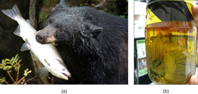
Even though members of the animal kingdom are incredibly diverse, most animals share certain features that distinguish them from organisms in other kingdoms. All animals are eukaryotic, multicellular organisms, and almost all animals have a complex tissue structure with differentiated and specialized tissues. Most animals are motile, at least during certain life stages. All animals require a source of food and are therefore heterotrophic, ingesting other living or dead organisms; this feature distinguishes them from autotrophic organisms, such as most plants, which synthesize their own nutrients through photosynthesis. As heterotrophs, animals may be carnivores, herbivores, omnivores, or parasites (Figureab). Most animals reproduce sexually, and the offspring pass through a series of developmental stages that establish a determined and fixed body plan. The body plan refers to the morphology of an animal, determined by developmental cues.

Complex Tissue Structure
As multicellular organisms, animals differ from plants and fungi because their cells don’t have cell walls, their cells may be embedded in an extracellular matrix (such as bone, skin, or connective tissue), and their cells have unique structures for intercellular communication (such as gap junctions). In addition, animals possess unique tissues, absent in fungi and plants, which allow coordination (nerve tissue) of motility (muscle tissue). Animals are also characterized by specialized connective tissues that provide structural support for cells and organs. This connective tissue constitutes the extracellular surroundings of cells and is made up of organic and inorganic materials. In vertebrates, bone tissue is a type of connective tissue that supports the entire body structure. The complex bodies and activities of vertebrates demand such supportive tissues. Epithelial tissues cover, line, protect, and secrete. Epithelial tissues include the epidermis of the integument, the lining of the digestive tract and trachea, and make up the ducts of the liver and glands of advanced animals.
The animal kingdom is divided into Parazoa (sponges) and Eumetazoa (all other animals). As very simple animals, the organisms in group Parazoa (“beside animal”) do not contain true specialized tissues; although they do possess specialized cells that perform different functions, those cells are not organized into tissues. These organisms are considered animals since they lack the ability to make their own food. Animals with true tissues are in the group Eumetazoa (“true animals”). When we think of animals, we usually think of Eumetazoans, since most animals fall into this category.
The different types of tissues in true animals are responsible for carrying out specific functions for the organism. This differentiation and specialization of tissues is part of what allows for such incredible animal diversity. For example, the evolution of nerve tissues and muscle tissues has resulted in animals’ unique ability to rapidly sense and respond to changes in their environment. This allows animals to survive in environments where they must compete with other species to meet their nutritional demands.
Link to Learning

Watch a presentation by biologist E.O. Wilson on the importance of diversity.
Animal Reproduction and Development
Most animals are diploid organisms, meaning that their body (somatic) cells are diploid and haploid reproductive (gamete) cells are produced through meiosis. Some exceptions exist: For example, in bees, wasps, and ants, the male is haploid because it develops from unfertilized eggs. Most animals undergo sexual reproduction: This fact distinguishes animals from fungi, protists, and bacteria, where asexual reproduction is common or exclusive. However, a few groups, such as cnidarians, flatworm, and roundworms, undergo asexual reproduction, although nearly all of those animals also have a sexual phase to their life cycle.
Processes of Animal Reproduction and Embryonic Development
During sexual reproduction, the haploid gametes of the male and female individuals of a species combine in a process called fertilization. Typically, the small, motile male sperm fertilizes the much larger, sessile female egg. This process produces a diploid fertilized egg called a zygote.
Some animal species—including sea stars and sea anemones, as well as some insects, reptiles, and fish—are capable of asexual reproduction. The most common forms of asexual reproduction for stationary aquatic animals include budding and fragmentation, where part of a parent individual can separate and grow into a new individual. In contrast, a form of asexual reproduction found in certain insects and vertebrates is called parthenogenesis (or “virgin beginning”), where unfertilized eggs can develop into new male offspring. This type of parthenogenesis is called haplodiploidy. These types of asexual reproduction produce genetically identical offspring, which is disadvantageous from the perspective of evolutionary adaptability because of the potential buildup of deleterious mutations. However, for animals that are limited in their capacity to attract mates, asexual reproduction can ensure genetic propagation.
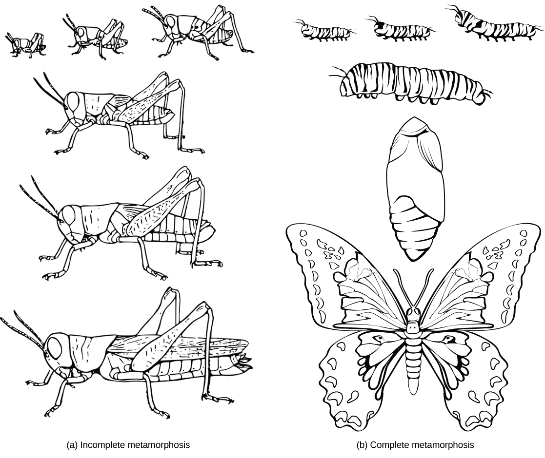
After fertilization, a series of developmental stages occur during which primary germ layers are established and reorganize to form an embryo. During this process, animal tissues begin to specialize and organize into organs and organ systems, determining their future morphology and physiology. Some animals, such as grasshoppers, undergo incomplete metamorphosis, in which the young resemble the adult. Other animals, such as some insects, undergo complete metamorphosis where individuals enter one or more larval stages that may in differ in structure and function from the adult (Figure). For the latter, the young and the adult may have different diets, limiting competition for food between them. Regardless of whether a species undergoes complete or incomplete metamorphosis, the series of developmental stages of the embryo remains largely the same for most members of the animal kingdom.
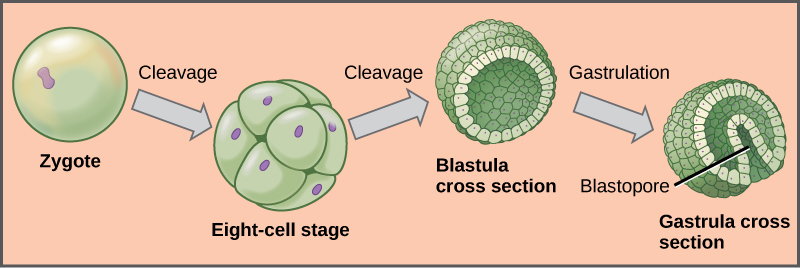
The process of animal development begins with the cleavage, or series of mitotic cell divisions, of the zygote (Figure). Three cell divisions transform the single-celled zygote into an eight-celled structure. After further cell division and rearrangement of existing cells, a 6–32-celled hollow structure called a blastula is formed. Next, the blastula undergoes further cell division and cellular rearrangement during a process called gastrulation. This leads to the formation of the next developmental stage, the gastrula, in which the future digestive cavity is formed. Different cell layers (called germ layers) are formed during gastrulation. These germ layers are programmed to develop into certain tissue types, organs, and organ systems during a process called organogenesis.
Link to Learning

Watch the following video to see how human embryonic development (after the blastula and gastrula stages of development) reflects evolution.
The Role of Homeobox (Hox) Genes in Animal Development
Since the early 19th century, scientists have observed that many animals, from the very simple to the complex, shared similar embryonic morphology and development. Surprisingly, a human embryo and a frog embryo, at a certain stage of embryonic development, look remarkably alike. For a long time, scientists did not understand why so many animal species looked similar during embryonic development but were very different as adults. They wondered what dictated the developmental direction that a fly, mouse, frog, or human embryo would take. Near the end of the 20th century, a particular class of genes was discovered that had this very job. These genes that determine animal structure are called “homeotic genes,” and they contain DNA sequences called homeoboxes. The animal genes containing homeobox sequences are specifically referred to as Hox genes. This family of genes is responsible for determining the general body plan, such as the number of body segments of an animal, the number and placement of appendages, and animal head-tail directionality. The first Hox genes to be sequenced were those from the fruit fly (Drosophila melanogaster). A single Hox mutation in the fruit fly can result in an extra pair of wings or even appendages growing from the “wrong” body part.
While there are a great many genes that play roles in the morphological development of an animal, what makes Hox genes so powerful is that they serve as master control genes that can turn on or off large numbers of other genes. Hox genes do this by coding transcription factors that control the expression of numerous other genes. Hox genes are homologous in the animal kingdom, that is, the genetic sequences of Hox genes and their positions on chromosomes are remarkably similar across most animals because of their presence in a common ancestor, from worms to flies, mice, and humans (Figure). One of the contributions to increased animal body complexity is that Hox genes have undergone at least two duplication events during animal evolution, with the additional genes allowing for more complex body types to evolve.
Art Connection
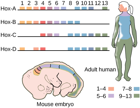
If a Hox 13 gene in a mouse was replaced with a Hox 1 gene, how might this alter animal development?
Section Summary
Animals constitute an incredibly diverse kingdom of organisms. Although animals range in complexity from simple sea sponges to human beings, most members of the animal kingdom share certain features. Animals are eukaryotic, multicellular, heterotrophic organisms that ingest their food and usually develop into motile creatures with a fixed body plan. A major characteristic unique to the animal kingdom is the presence of differentiated tissues, such as nerve, muscle, and connective tissues, which are specialized to perform specific functions. Most animals undergo sexual reproduction, leading to a series of developmental embryonic stages that are relatively similar across the animal kingdom. A class of transcriptional control genes called Hox genes directs the organization of the major animal body plans, and these genes are strongly homologous across the animal kingdom.
Art Connections
Review Questions
Which of the following is not a feature common to most animals?
- development into a fixed body plan
- asexual reproduction
- specialized tissues
- heterotrophic nutrient sourcing
Hint:
B
During embryonic development, unique cell layers develop and distinguish during a stage called ________.
- the blastula stage
- the germ layer stage
- the gastrula stage
- the organogenesis stage
Hint:
C
Which of the following phenotypes would most likely be the result of a Hox gene mutation?
- abnormal body length or height
- two different eye colors
- the contraction of a genetic illness
- two fewer appendages than normal
Hint:
D
Free Response
Why might the evolution of specialized tissues be important for animal function and complexity?
Hint:
The development of specialized tissues affords more complex animal anatomy and physiology because differentiated tissue types can perform unique functions and work together in tandem to allow the animal to perform more functions. For example, specialized muscle tissue allows directed and efficient movement, and specialized nervous tissue allows for multiple sensory modalities as well as the ability to respond to various sensory information; these functions are not necessarily available to other non-animal organisms.
Describe and give examples of how humans display all of the features common to the animal kingdom.
Hint:
Humans are multicellular organisms. They also contain differentiated tissues, such as epithelial, muscle, and nervous tissue, as well as specialized organs and organ systems. As heterotrophs, humans cannot produce their own nutrients and must obtain them by ingesting other organisms, such as plants, fungi, and animals. Humans undergo sexual reproduction, as well as the same embryonic developmental stages as other animals, which eventually lead to a fixed and motile body plan controlled in large part by Hox genes.
How have Hox genes contributed to the diversity of animal body plans?
Hint:
Altered expression of homeotic genes can lead to major changes in the morphology of the individual. Hox genes can affect the spatial arrangements of organs and body parts. If a Hox gene was mutated or duplicated, it could affect where a leg might be on a fruit fly or how far apart a person’s fingers are.