Unrestricted Use
CC BY
- Subject:
- Anatomy/Physiology
- Material Type:
- Module
- Author:
- Mindy Boland
- Date Added:
- 02/18/2020

This set of anatomy videos illustrating parts of the human body was created under a Round Eleven Mini-Grant for Ancillary Materials Creation.
Topics include:
Axial Skeleton
Appendicular Skeleton
Muscles
Nervous System
Anatomy of the Senses

Short, animated videos on many Anatomy and Physiology topics. Videos used in college courses and cover the content presented in the first 2 semesters of Anatomy and Physiology for Nursing/Allied Health students.
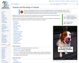
Veterinary nurses need to have a firm grasp of the normal structure of an animal’s body and how it functions before they can understand the effect diseases and injuries have and the best ways to treat them. This book describes the structure of the animal body and the way in which it works. Animals encountered in normal veterinary practice are used as examples where possible.
Lab 1: Overview & The Microscope
Lab 2: Cytology
Lab 3: Histology
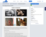
Lab 4: The Integumentary System
Lab 5: The Axial Skeleton
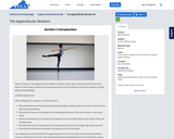
Lab 6: The Appendicular Skeleton
Lab 7: Joints
Lab 8: The Axial Muscles
Lab 9: The Appendicular Muscles
Lab 10: Nervous Tissue
Lab 11: The Central Nervous System (Brain)
Lab 12: Cranial and Spinal Nerves
Lab 13: The Somatic Nervous System (Special Senses)
Lab 14: The Endocrine System
Lab 15: Blood
Lab 16: The Heart
Lab 17: Blood Vessels and Circulation
Lab 18: The Lymphatic System
Lab 19: The Respiratory System
Lab 20: The Digestive System
Lab 21: The Urinary System
Lab 22: The Reproductive System (Male)
Lab 23: The Reproductive System (Female)

This course is an introduction to the moral challenges that arise in the design and execution of biomedical research and the development of medical interventions. A historical review segues into detailed examination of key ethical concepts and principles, as well as topics of particular concern. At the culmination of the semester, students apply their knowledge of research ethics to an ethical analysis of their MTM BioDesign projects.
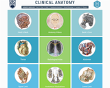
Interactive radiology images, animated modules showing the physiology of difficult to understand muscle groups, sketches of anatomy, and links to the already existing quality neuroanatomy website.
Covers:
Head and Neck
Anatomy Videos
Back and Core
Thorax
Radiological Atlas
Abdomen
Upper Limb
Anatomical Illustrations
Lower Limb
Embryology
Neuroanatomy
Pelvis
This animation describes Endochondral Ossification which is a process of long, short and irregular bone formation using byline template.
This animation describes the formation of endocrine glands starting with the mitosis of mesenchymal tissues, eventually leading to the formation of the secretory portion of the gland by differentiation.
The OER content in this course shell was originally intended for a 300-level Kinesiology & Biomechanics course. The course is described as providing "an in-depth study of the basic body movements, osteology, applied myology, spatial relations of muscles and joints, aggregate muscle action, kinesiologic constructs of summation of internal forces, aerodynamics and hydrodynamics, techniques for cinematographical and noncinematographical analysis of sport skills. The study of methods, mechanics and analysis of movement as applied to the structure and function of the human organism will also be discussed"
This book is a guide to the basic fetal pig dissection conducted as a part of the Queens College, CUNY Biology Department Bio105 General Biology: Physiology and Cell Biology course. This course is the first half our two-part series for biology majors. The actives are designed to be conducted over a three- 3-hour lab periods which focus on the relationship of form and function of the pig anatomy and physiology. Step by step instructions for the dissection are provided along with some microscopy tasks to look at the histology of key organs.
In addition to the full text of the book, we also provide a form with just the assessment portions of the book. This allows students to limit the printed material to just those pages.
This animation describes the process of cartilage formation starting with separation from the mesenchymal tissue to the formation of an isogenous cell group via mitosis.
Short Description:
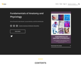
Fundamentals of Anatomy and Physiology is a textbook for biomedical, life science and health majors. The book is organised by body system and contains interactive resources to test your knowledge.
Word Count: 416894
ISBN: 978-0-6487698-5-9
(Note: This resource's metadata has been created automatically by reformatting and/or combining the information that the author initially provided as part of a bulk import process.)
Quiz questions that accompany the text are available for faculty and instructors. Request access by providing your credentials and contacting us at learnlib@umn.edu.
This book is a guide to the basic histology lab conducted as a part of the Queens College, CUNY Biology Department Bio105 General Biology: Physiology and Cell Biology course. This course is the first half our two-part series for biology majors. The actives are designed to be conducted over a single 3-hour lab periods which focus on the relationship of form and function of the cellular and organ level anatomy and physiology. Step by step instructions for each slide set are provided for all the key organs.
In addition to the full text of the book, we also provide a checklist form with just the assessment portions of the book. This is to help summarize all the information the student should get from the activity.
Human biology is an interdisciplinary area of study that examines humans through the influences and interplay of many diverse fields such as genetics, evolution, physiology, anatomy, epidemiology, anthropology, ecology, nutrition, population genetics, and sociocultural influences.
Short Description:
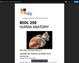
This is a lab manual for a college-level human anatomy course. Mastery of anatomy requires a fair amount of memorization and recall skills. The activities in this manual encourage students to engage with new vocabulary in many ways, including grouping key terms, matching terms to structures, recalling definitions, and written exercises. Most of the activities in this manual utilize anatomical models, and several dissections of animal tissues and histological examinations are also included. Each unit includes both pre- and post-lab questions and six lab exercises designed for a classroom where students move from station to station. The vocabulary terms used in each unit are listed at the end of the manual and serve as a checklist for practicals.
Word Count: 24242
(Note: This resource's metadata has been created automatically by reformatting and/or combining the information that the author initially provided as part of a bulk import process.)
This human anatomy laboratory manual acts as a textbook for undergraduate human anatomy courses. Each chapter has review questions and laboratory activities, and most chapters also have collaborative learning activities. There are 22 chapters total. The chapters are:
Chapter 1: Introduction to Anatomy & Anatomical Terms
Chapter 2: Introduction to Microscopes
Chapter 3: Cell Structures & Types
Chapter 4: How Cells Divide (Mitotic Cell Division)
Chapter 5: Tissues
Chapter 6: Integumentary System
Chapter 7: Introduction to the Skeletal System
Chapter 8: Axial Skeleton
Chapter 9: Appendicular Skeleton
Chapter 10: Articulations (Joints) & Movements
Chapter 11: Introduction to Skeletal Muscles
Chapter 12: The Skeletal Muscles
Chapter 13: Introduction to the Nervous System
Chapter 14: Central Nervous System
Chapter 15: Peripheral Nervous System
Chapter 16: Special Senses of the Nervous System
Chapter 17: Cardiovascular System - The Heart
Chapter 18: Cardiovascular System - The Blood Vessels
Chapter 19: Respiratory System
Chapter 20: Digestive System
Chapter 21: Urinary System
Chapter 22: Reproductive Systems